

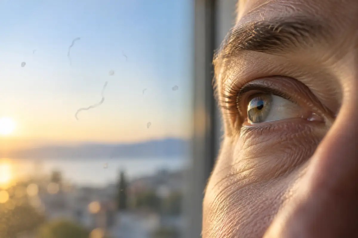
Almost everyone has at some point noticed tiny dots or threads “floating” in front of their eyes while looking at a clear sky or a white wall. Such floaters are mostly harmless. However, when they appear suddenly, in large numbers or with flashes of light, they can be the first sign of retinal detachment – a condition that requires a prompt examination. The good news is that with timely treatment vision can be saved in most cases, and the key is to recognise the warning signs in time.
What is the retina and why can it detach?
The retina is a thin layer of nerve tissue on the back wall of the eye that turns light into signals for the brain. The inside of the eye is filled with the vitreous, a clear gel-like substance. With age it becomes more liquid and shrinks, so at some point it separates from the retina. This process is called posterior vitreous detachment and is very common after the age of fifty.
Most often it causes no harm. Sometimes, however, the vitreous pulls on the retina as it separates and makes a small tear. Fluid can pass through it under the retina and lift it from its base – this is called retinal detachment. According to a large study reported by the American Academy of Ophthalmology, a retinal tear was found in about 5% of patients with vitreous symptoms, which shows why every new symptom should be checked.
What do the first signs look like?
Retinal detachment usually doesn’t hurt, which makes it easy to ignore. According to the US National Eye Institute (NEI) i UK’s NHS, the most important signs are:
- sudden appearance of new floaters or a marked increase in their number (a “shower” of dots),
- flashes of light, especially at the edge of the visual field or in the dark,
- a dark shadow or “curtain” spreading from the edge towards the centre of the visual field,
- suddenly blurred or distorted vision.
Symptoms usually occur in one eye. If you cover one eye with your palm and compare your vision, you will notice the change more easily.
Who is at higher risk?
Retinal detachment can happen to anyone, but the risk is higher in:
- people over 50,
- short-sighted people, especially with a high prescription,
- people who have had eye surgery, for example cataract surgery,
- people after a serious eye injury,
- those who have already had a retinal detachment in the other eye or have a family history of it,
- people with diabetic retinopathy or conditions that thin the retina.
Belonging to one of these groups doesn’t mean your retina will detach, but it is useful to know the symptoms and have regular examinations.
How is it diagnosed and treated?
The ophthalmologist first dilates the pupil with drops and carefully examines the whole back of the eye, including the peripheral retina, where tears most often occur. Optical coherence tomography (OCT) imaging helps assess the central retina. Such a thorough examination is part of our Premium Plus eye examination.
If only a tear is found, it is often enough to seal it with a laser or freezing treatment, which prevents further detachment. When the retina has already detached, surgery is needed. According to the NEI, treatment is successful in about 9 out of 10 people, and the outcome is better the earlier you act.
When to see an ophthalmologist?
See an ophthalmologist the same day if you notice a sudden appearance of floaters, flashes of light, a shadow or curtain in your field of vision, or a sudden drop in vision. If a same-day examination isn’t possible, go to an emergency eye service. Don’t wait for the symptoms to “go away on their own” – even when floaters decrease over time, a retinal tear can remain.
If you have long been used to a few unchanging floaters, that is usually not a reason for urgency, but it is worth mentioning them at your next specialist eye examination.
Conclusion
Floaters are mostly a harmless part of the ageing eye, but a sudden change, flashes or a shadow in your field of vision are signs that the eye needs urgent examination. A quick reaction is the best protection for your vision. If you have noticed new symptoms or want to check the condition of your retina, call us at 047 655 999 or contact us via our contact page. At Poliklinika Pavan we will be happy to examine you.
Sources: National Eye Institute (NEI) – Retinal Detachment; NHS – Detached retina; American Academy of Ophthalmology, EyeNet – Rates and predictors of PVD.